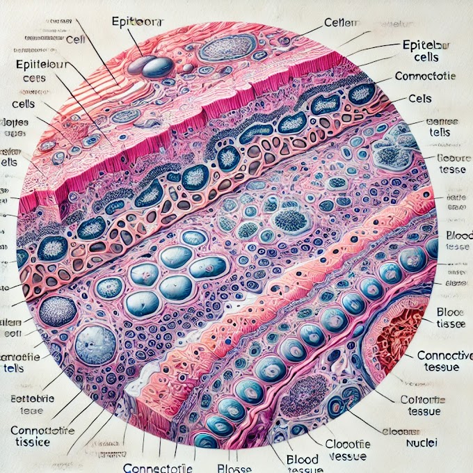
Identifying peripheral nerve tissue on histology slides involves recognizing specific features unique to nerves. Here are key identification points for peripheral nerve histology:
Epineurium, Perineurium, and Endoneurium:
Nerves are organized into fascicles, bundles of nerve fibers, surrounded by connective tissue.
The epineurium surrounds the entire nerve.
The perineurium surrounds each fascicle.
The endoneurium surrounds individual nerve fibers.
Axons:
Nerve fibers (axons) are the main cellular components of nerves.
Axons are often arranged in bundles within the fascicles.
Myelin Sheath:
Myelinated axons are surrounded by a myelin sheath, which appears as a lighter-staining region.
Unmyelinated axons lack this distinct myelin sheath.
Nodes of Ranvier:
Nodes of Ranvier are periodic gaps in the myelin sheath along myelinated axons.
They play a crucial role in the conduction of nerve impulses.
Schwann Cells:
Schwann cells are glial cells that produce the myelin sheath around peripheral nerve axons.
Each Schwann cell myelinates a segment of a single axon.
Blood Vessels:
Nerves are vascularized, and blood vessels can be seen in the epineurium.
Adequate blood supply is essential for the metabolic needs of nerve tissue.
Connective Tissue Layers:
The connective tissue layers, including the epineurium, perineurium, and endoneurium, provide structural support and protection to the nerve fibers.
Staining Characteristics:
Standard histological stains, such as hematoxylin and eosin (H&E), can be used to visualize the cellular components and connective tissue of nerves.
Cross-Sectional and Longitudinal Views:
Depending on the orientation of the tissue section, nerves may be viewed in cross-section or longitudinally.
Cross-sectional views reveal the circular arrangement of fascicles.
Neuromuscular Junctions (if present):
In motor nerves that innervate muscles, the neuromuscular junctions may be visible where the nerve fibers synapse with muscle fibers.
overview of the anatomy, physiology, histology, histopathology, and clinical significance of peripheral nerves:
1. Anatomy
Peripheral nerves consist of bundles of nerve fibers (axons) organized into fascicles and encased by connective tissue layers:
Epineurium: The outermost layer surrounding the entire nerve, providing protection and structural support.
Perineurium: The connective tissue surrounding each fascicle (bundle of axons), which maintains the blood-nerve barrier.
Endoneurium: The delicate layer around individual axons, providing insulation and supporting metabolic exchange.
Axons within each fascicle can be myelinated or unmyelinated, with myelinated fibers conducting signals faster due to the insulating myelin sheath produced by Schwann cells.
2. Physiology
Peripheral nerves conduct electrical signals from the central nervous system to peripheral organs, muscles, and skin and bring sensory information back to the CNS. They contain motor (efferent) fibers that control movement and sensory (afferent) fibers that transmit sensations like touch, pain, and temperature.
Myelin sheaths around certain axons increase the speed of signal transmission, allowing rapid response to stimuli.
3. Histology
Under a microscope, peripheral nerve sections reveal:
Fascicles: Clearly visible bundles of axons, each surrounded by the perineurium.
Connective Tissue Layers: The epineurium, perineurium, and endoneurium can be seen encasing the nerve structure.
Myelinated vs. Unmyelinated Axons: Myelinated axons appear larger due to the myelin sheath, while unmyelinated axons are smaller and less distinct.
Staining techniques like hematoxylin and eosin (H&E) or special nerve stains (e.g., Luxol fast blue for myelin) help differentiate these layers and structures.
4. Histopathology
Peripheral nerves can be affected by a range of pathological conditions:
Neuropathy: Caused by diabetes, toxins, or trauma, resulting in axonal degeneration or demyelination, which impairs signal conduction.
Inflammation (Neuritis): Conditions like Guillain-Barré syndrome involve immune-mediated attack on peripheral nerves, leading to demyelination.
Tumors (Neurofibromas, Schwannomas): These tumors arise from nerve sheath cells and may affect nearby nerves, causing pain or dysfunction.
Trauma: Injuries can result in nerve transection or crush injuries, leading to Wallerian degeneration, where axons distal to the injury site degenerate.
5. Clinical Significance
Peripheral nerves are essential for both voluntary and involuntary functions. Damage can result in:
Motor Deficits: Weakness or paralysis due to motor nerve involvement.
Sensory Deficits: Numbness, tingling, or pain from sensory nerve damage.
Autonomic Dysfunction: Certain peripheral nerves control autonomic functions like sweating or blood vessel regulation, and damage may affect these systems.
Clinical conditions like diabetic neuropathy, carpal tunnel syndrome, and peripheral neuropathy have significant impacts on patients' quality of life, requiring diagnostic techniques like nerve conduction studies and treatment options including medication, physical therapy, and sometimes surgery.
Written By: Ikrambaigtech
click here to wach all nervous tissue slides videos on my youtube channel ikrambiag@tech


0 Comments