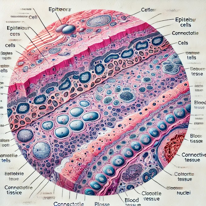
Under The Light Microscopic View
Empty-Looking Cells (Adipocytes):
Adipocytes are the essential cells in fat tissue. They show up as huge, round or oval cells with an unmistakable or void looking cytoplasm.
Signet Ring Appearance:
Connective Tissue Septa:
Ovoid Closely Packed Cells:
Marginal Nuclei:
Cellular Composition:
- Adipose tissue is primarily composed of adipocytes, which are specialized cells for storing fat.
- The nucleus of an adipocyte is typically pushed to the periphery of the cell due to the large fat droplet in the center.
Unilocular Fat Cells:
- Adipocytes in white adipose tissue are unilocular, meaning they have a single, large fat droplet in the cell.
- This single lipid droplet occupies most of the cell volume, displacing the nucleus and other organelles to the periphery.
Cytoplasm and Nucleus:
- The cytoplasm of an adipocyte is a thin rim around the large fat droplet.
- The nucleus appears flattened and pushed to the edge of the cell by the fat droplet.
Lack of Matrix:
- Adipose tissue has a minimal extracellular matrix compared to other connective tissues.
- The cells are loosely arranged and separated by a delicate network of reticular fibers.
Blood Vessels:
- Adipose tissue is vascularized, and blood vessels are present between adipocytes.
- The blood vessels supply nutrients and oxygen to the adipose tissue.
Staining Characteristics:
- Adipose tissue stains well with lipophilic stains, such as Sudan dyes or Oil Red O, which highlight the presence of lipid droplets.
Distribution:
- Adipose tissue is found throughout the body, with varying distributions, including subcutaneous adipose tissue beneath the skin and visceral adipose tissue surrounding organs.
Functions:
- Adipose tissue serves as an energy storage site and provides insulation and cushioning for organs.
- Brown adipose tissue, which is involved in thermogenesis, has a distinct appearance and function compared to white adipose tissue.
Histology Slide of Adipose Connective Tissue
Features:
Adipocytes (Fat Cells):
- Large Lipid Droplets: The adipocytes are characterized by large, round lipid droplets that occupy most of the cell volume. These droplets appear white or clear because the lipid content is often removed during the preparation of the slide.
- Cell Membranes: The cell membranes of adipocytes are thin and delicate, surrounding the large lipid droplet. The nucleus and cytoplasm are pushed to the periphery of the cell, creating a signet ring appearance.
- Shape and Size: Adipocytes are relatively large compared to other cell types and have a spherical or oval shape.
Connective Tissue Fibers:
- Collagen Fibers: These fibers provide structural support to the adipose tissue and appear as thin, wavy lines interspersed between adipocytes. They stain pink with hematoxylin and eosin (H&E) staining.
- Elastic Fibers: Though not always prominent, elastic fibers may be present and contribute to the tissue's flexibility.
Blood Vessels:
- Capillaries and Small Blood Vessels: The adipose tissue is richly vascularized, with numerous small blood vessels running through it. These vessels are crucial for supplying nutrients and oxygen to the adipocytes and for removing metabolic waste products.
- Endothelial Cells: The lining of these blood vessels is composed of endothelial cells, which are visible as a thin layer around the vessel lumen.
Extracellular Matrix:
- Ground Substance: The extracellular matrix includes a ground substance that fills the spaces between cells and fibers. This substance provides support and facilitates nutrient and waste exchange.
Fibroblasts:
- Support Cells: Fibroblasts are responsible for producing and maintaining the extracellular matrix and connective tissue fibers. They are less prominent than adipocytes but can be seen as elongated cells within the tissue.
Best 3 Identification Points for Medical Students:
Signet Ring Appearance of Adipocytes:
- The most distinctive feature of adipose tissue is the signet ring appearance of adipocytes. The large central lipid droplet displaces the nucleus to the cell's periphery, creating a characteristic ring shape. This is a key point for identifying adipose tissue under the microscope.
Rich Vascular Network:
- The presence of numerous capillaries and small blood vessels within the adipose tissue is another important identification feature. The rich vascular network supports the metabolic activity of the adipocytes and is essential for the tissue's function.
Thin Connective Tissue Fibers:
- Adipose tissue contains a delicate network of collagen fibers that provide structural support. These fibers are visible as thin, pink-stained lines running between the adipocytes. Identifying these fibers helps distinguish adipose tissue from other types of connective tissue.
This description and these identification points provide medical students with a comprehensive understanding of the histological features of adipose connective tissue, aiding in their studies and practical examinations.
Overview of Adipose Tissue
Anatomy
Adipose tissue, commonly known as body fat, is a type of connective tissue that stores energy in the form of lipids. It is primarily composed of adipocytes (fat cells) and is found throughout the body. There are two main types of adipose tissue:
- White Adipose Tissue (WAT): The most prevalent type, WAT is responsible for storing energy and acting as an insulator and cushion for organs.
- Brown Adipose Tissue (BAT): Found in smaller quantities, BAT generates heat and plays a role in thermoregulation, particularly in infants and hibernating animals.
Physiology
Adipose tissue serves several crucial functions:
- Energy Storage: Adipocytes store triglycerides, which can be broken down into fatty acids and glycerol to provide energy during periods of fasting or increased energy demand.
- Insulation and Protection: Fat acts as an insulator, helping to maintain body temperature, and cushions organs against mechanical shock.
- Endocrine Function: Adipose tissue secretes various hormones and cytokines (adipokines), such as leptin, adiponectin, and resistin, which regulate appetite, metabolism, and inflammation.
Biology
Adipose tissue is a dynamic organ that can expand or shrink in response to energy balance. Adipocytes can increase in size (hypertrophy) or number (hyperplasia) depending on nutritional status. The tissue is also highly vascularized and innervated, which is essential for its metabolic activity.
Histopathology
Histopathological examination of adipose tissue can reveal various conditions and abnormalities:
- Obesity: Characterized by an increase in the size and number of adipocytes, leading to excessive fat accumulation.
- Lipodystrophy: A condition where there is abnormal distribution of adipose tissue, either as loss (lipoatrophy) or abnormal accumulation.
- Inflammation: In obesity, adipose tissue can become infiltrated with immune cells, leading to chronic inflammation, which is linked to insulin resistance and metabolic syndrome.
- Tumors: Adipose tissue can develop benign tumors such as lipomas or malignant tumors like liposarcomas.
Clinical Significance
Adipose tissue plays a significant role in various health conditions:
- Obesity: Excess adipose tissue is a major risk factor for cardiovascular disease, type 2 diabetes, and certain cancers. Managing obesity involves lifestyle changes, medications, and sometimes surgery.
- Metabolic Syndrome: A cluster of conditions (increased blood pressure, high blood sugar, excess body fat around the waist, and abnormal cholesterol levels) that occur together, increasing the risk of heart disease, stroke, and diabetes.
- Diabetes: Excess adipose tissue, particularly visceral fat, is associated with insulin resistance and type 2 diabetes.
- Liposuction: A cosmetic procedure to remove excess fat from specific areas of the body.
- Thermogenesis and Weight Loss: BAT plays a role in energy expenditure and thermogenesis, making it a target for obesity treatment. Strategies to activate BAT include exposure to cold and certain medications.
Conclusion
Adipose tissue is a vital component of the human body, with diverse functions ranging from energy storage to hormone secretion. Understanding its anatomy, physiology, biology, histopathology, and clinical significance is essential for addressing various health conditions related to adipose tissue. Advances in research continue to uncover new aspects of adipose tissue function and its role in health and disease.
written by: ikrambaigtech
diagram Of C.T


%20with%20clear%20cell%20membranes,%20large%20.webp)


%20Volume.webp)


0 Comments